Hafiz Muhammad Irfan1,2 ![]() ,
Mohd Zaini Asmawi1,
Nurzalina Abdul Karim Khan1,
Amirin Sadikun1,
Mohd Nizam Mordi3
,
Mohd Zaini Asmawi1,
Nurzalina Abdul Karim Khan1,
Amirin Sadikun1,
Mohd Nizam Mordi3
For correspondence:- Hafiz Irfan Email: irfan_pharmacist06@yahoo.com Tel:+60104300950
Received: 3 October 2016 Accepted: 8 February 2017 Published: 31 March 2017
Citation: Irfan HM, Asmawi M, Khan NA, Sadikun A, Mordi MN. Anti-diabetic activity-guided screening of aqueous-ethanol Moringa oleifera extracts and fractions: Identification of marker compounds. Trop J Pharm Res 2017; 16(3):543-552 doi: 10.4314/tjpr.v16i3.7
© 2017 The authors.
This is an Open Access article that uses a funding model which does not charge readers or their institutions for access and distributed under the terms of the Creative Commons Attribution License (http://creativecommons.org/licenses/by/4.0) and the Budapest Open Access Initiative (http://www.budapestopenaccessinitiative.org/read), which permit unrestricted use, distribution, and reproduction in any medium, provided the original work is properly credited..
Purpose: To explore the anti-diabetic effects of Moringa oleifera extracts and fractions, and to identify their active/marker compounds.
Methods: Five different aqueous ethanol extracts (95, 75, 50, 25 %v/v and 100 % water) of Moringa oleifera were given orally to normal rats to assess their hypoglycemic activities and effect on intraperitoneal glucose tolerance test (IPGTT) data. Rats with streptozotocin-induced diabetes were used to assess acute and sub-chronic anti-hyperglycemic activities. The most active extract was further subjected to liquid-liquid fractionation into hexane, chloroform, ethyl acetate, butanol, and water; these fractions were screened for anti-diabetic activities. The most active extract, and fractions thereof, were then subjected to qualitative and quantitative phytochemical analysis. Standardization was achieved via thin layer chromatography (TLC) and high-performance liquid chromatography (HPLC), and used to identify marker compounds.
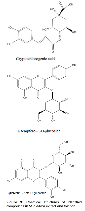
Results: Of all the extracts and fractions, 95 % (v/v) ethanol extract (at 1,000 mg/kg) and the butanol fraction thereof (at 500 mg/kg) were the most active, reducing blood glucose concentration after one-time (acute) administration to diabetic rats (p < 0.01). No significant hypoglycemic activity was apparent, and the materials had no effect on IPGTT performance by normal rats. TLC and HPLC identified quercetin 3-β-D-glucoside, kaempferol-3-O-glucoside, and cryptochlorogenic acid.
Conclusion: An M. oleifera leaf extract exhibited anti-hyperglycaemic activity in diabetic rats only. This effect was likely attributable to cryptochlorogenic acid, quercetin 3-β-D-glucoside, and kaempferol 3-O-glucoside
Introduction
Glucose homeostasis is the balance between glucose production and utilisation by peripheral tissues. Insulin promotes glucose uptake by cells [1]. An insulin deficiency combined with insulin resistance is responsible for diabetes mellitus [2]. The World Health Organization has observed that diabetes has become a major problem, and the number of cases is expected to rise through the year 2035 [3]. The prevalence of diabetes is increasing among Malaysians, reaching 23.8 % in recent years [4].
Natural products and their derivatives constitute ca. 50 % of all anti-diabetics. The rate is higher when related synthetic materials are considered [5], but these materials are costly [6]. Generally, various extracts of natural materials are subjected to primary screening and the best extract is selected [7].
Marker compounds therein are identified using standard techniques [8]. High-performance liquid chromatography (HPLC) and thin layer chromatography (TLC) are valuable chromatographic techniques [9] employed to identify molecules responsible for therapeutic activities [10]. Recently, advances in the isolation and identification of active compounds in medicinal plants have featured fractionation-guided targeting.
Therefore, we identified active anti-diabetic compounds in extracts and fractions of Moringa oleifera. Our findings will aid future drug discovery/extraction from natural sources.
Methods
Apparatus, chemicals, and drugs
A rotary evaporator (Buchi Labotechnik, CH-9230 Flawil Switzerland), a WinCATS Planar Chromatography Manager (Merck, Germany), streptozotocin (Sigma Aldrich Chemical Co, USA), acetonitrile and formic acid (Merck, Germany), quercetin 3-β-D-glucoside (Sigma Aldrich, USA), kaempferol 3-O-glucoside (480-10-4, Extrasynthese, Genay Cedex), cryptochlorogenic acid (Wuhan ChemFaces Biochemical Co. Ltd), glibenclamide 5 mg (Daonil) and metformine (500 mg; Glucophage; Merck, Germany) were used in this study.
Authentication of the plant and preparation of extracts
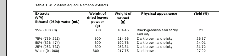
Plant material was purchased from Harbagus Sdn. Bhd., Pulau Pinang, Malaysia, and was authenticated by Dr Rahmad Zakaria. A voucher specimen has been placed in the herbarium of School of botany, Universiti Sains Malaysia (no. 11626). M. oleifera leaves were dried in the shade, ground to a powder, and extracted with 95, 75, 50, and 25 % (v/v) ethanol and 100 % water via maceration (). All extracts were concentrated under reduced pressure in a rotary evaporator and dried in an oven at 45 ºC.
Experimental animals
Healthy male adult Sprague Dawley (SD) rats aged 8 - 9 weeks (230 ± 30 g body weight) were used in the experimental studies. The animals were obtained from the Animal Research and Service Centre (ARASC), Universiti Sains Malaysia and allowed to adapt for seven days in the transit room of the School of Pharmaceutical Sciences at a temperature of 25 ± 3 °C under a 12/12-h dark/light cycle. The rats had ad libitum access to rat pellets and water. The Animal Ethics Committee of Universiti Sains Malaysia approved the work (approval no. AEA/2015/612) and was performed in compliance with international guidelines (NIH) for the care and use of animals [11].
Blood glucose measurement
Blood samples (single drops) were collected by nicking the tail vein, and blood glucose (BG) levels were determined using a glucometer (Roche, ACCU-CHEK Performa no. 55404068967).
Anti-diabetic studies
Hypoglycaemia in normoglycaemic rats
Forty-two male SD rats were used to measure hypoglycaemic activity. The negative control (NC) and positive control (PC) groups were given distilled water (10 mL/kg) and water containing glibenclamide (10 mg/kg), respectively. The treated groups were given 95, 75, 50 and 25 % (v/v) ethanol and 100 % water extracts (1,000 mg/kg orally). After 14 h overnight fasting, fasting blood glucose (FBG) levels were determined; the treatments were then applied, and glucose levels were measured 1, 3, 5 and 7 h later.
Intraperitoneal glucose tolerance test in normoglycaemic rats
Normal rats were divided randomly into eight groups. One hour after oral administration of all treatments, the rats were injected intraperitoneally with a glucose load (500 mg/kg body weight). BG levels were measured at 0, 15, 30, 45, 60, 90, and 120 min thereafter.
Anti-hyperglycaemic activity in streptozo-tocin-treated rats: single-dose study
Diabetes was induced by intraperitoneal injection of streptozotocin (45 mg/kg body weight) after overnight fasting. Glucose was added to the drinking water (to 5 %, w/v) for 12 h to reduce mortality caused by hypoglycaemia. BG levels were measured 72 h later. Rats with FBG concentrations > 15 mmol/L were considered to be diabetic and were used in further studies.
To measure anti-hyperglycaemic activity, rats were divided into seven groups. The experimental protocol was as described above, except that the PC group received metformin (500 mg/kg).
Anti-hyperglycaemic activity in streptozo-tocin-treated rats: multiple-dose study
The groups formed were the same as those created for the single-dose study described above. Diabetic rats were treated with M. oleifera aqueous ethanol extracts (1,000 mg/kg) once daily for 14 days. BG levels were determined on day-0, 7 and 14 after overnight fasting.
Fractionation (Liquid-liquid extraction)
The most active extract (the 95 % [v/v] ethanol) of M. oleifera was subjected to further fractionation. The extract was first partitioned with distilled water, and then (serially) with hexane, chloroform, ethyl acetate, and butanol, in a separating funnel. All five fractions (hexane, chloroform, ethyl acetate, butanol, and water) were concentrated separately under reduced pressure in rotary evaporator and dried in an oven at 45 °C.
Anti-hyperglycaemic screening of M. oleifera fractions in the rat streptozotocin model
Seven groups (six rats per group) were used. All groups underwent treatment with fractions (500 mg/kg) suspended in 10 % (v/v) Tween 80. FBG levels were determined after administration of various fractions and a standard drug at 1, 3, 5, and 7 h.
Phytochemical screening of extracts and fractions
For qualitative phytochemical screening, samples (extracts and fractions) were dissolved in dimethyl sulfoxide. Tannins, phenols, saponins, flavonoids, alkaloids, terpenoids, and glycosides were assayed using standard tests [12,13].
Thin layer chromatography
Qualitative screening of active compounds was performed on 20 × 20-cm silica gel TLC plates (Merck). The samples (the 95 % ethanol extract and the butanol fraction) were dissolved to 5 mg/mL and the standards (cryptochlorogenic acid, kaempferol-3-O-glucoside, and quercetin 3-β-D-glucoside) were dissolved to 1 mg/mL. The TLC plates were developed in a TLC chamber (10 × 10 cm); the mobile phase was ethyl acetate: methanol: Acetic acid: water (110: 13.5: 8: 8 by volume). The plates were sprayed with the natural product (NP) reagent and viewed under ultraviolet light at 254 and 366 nm with the aid of a winCATS Planar Chromatography Manager. Rf values were calculated using equation 1:
Rf = distance travelled by compound / distance travelled by solvent front ………... (1)
HPLC development and validation
A 95 % (v/v) ethanol extract, the butanol fraction thereof (1,000 ppm), and standard compound (2,000 ppm) stock solutions were prepared in methanol, filtered through syringe filter (0.45-µm pore size), and analysed on an 1100/1200 HPLC plateform (Agilent Technologies, USA) fitted with a column thermostat, a quaternary pump, a diode array detector, and an agilent 1200 autosampler (SL 1). We used a Hypersil Gold C4 column with a particle size of 5.0 µm and dimensions of 250.0 × 4.6 mm (Thermo-Scientific, USA). The solvent systems were 0.1 % (v/v) formic acid (B) and acetonitrile (C), and elution employed the following gradient: 0 - 5 min, B (97 %) and C (3 %); 5 - 14.5 min, B (80 %) and C (20 %); 14.5 - 16.5 min, B (97 %) and C (3 %). The injection volume was 20 µL, the column flow rate was 1.2 mL/min, and the run time was 18 min. All chromatographic signals were detected at 344 nm.
To quantify cryptochlorogenic acid, quercetin 3-β-D-glucoside, and kaempferol-3-O-glucoside in the active 95 % (v/v) ethanol extract and the butanol fraction thereof, the peak heights of samples were compared with those of standards.
Linearity was checked by evaluating the peak heights (in mAU) of standards (12.5 - 200 µg/mL). Accuracy was evaluated by checking agreement between the amounts applied and the amounts measured by HPLC. We also evaluated the repeatability (precision) of the method. We injected three sets of standard mixtures three times daily over 3 consecutive days and measured intra-day and inter-day accuracy and precision.
Statistical analysis
The results are presented as mean ± standard deviations and were analyzed by one-way analysis of variance with the aid of IBM SPSS software, followed by Dunnett’s post hoc test. Differences between means were considered to be statistically significant at p < 0.05, < 0.01, or < 0.001. For graphical presentation, GraphPad Prism (version 6.0) software was used.
Results
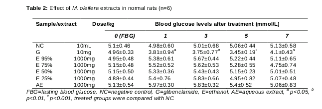
Effect of extracts of Moringa oleifera in normal rats
BG concentrations rose after administration of all extracts (95, 75, 50, 25 % [v/v] ethanol and water) for up to 3 h (compared with FBG levels), but declined over the next 4 h (). No significant reduction in glucose concentration was observed, except in the glibenclamide group (31.81 %).
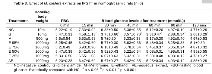
Effect of different extracts on IPGTT in normoglycemic rat
Glibenclamide greatly lowered the BG concentration (by 35.52 %) 15 min after glucose loading, compared with the NC. Similarly, the treated groups exhibited some improvements in glucose tolerance (GT) at 15 min, but these differences were not significant. The 95 and 50 % (v/v) ethanol extracts afforded only 12.41 % improvement in GT. GTs were 5.4, 10.64, and 11.73 % after administration of 75 and 25 % (v/v) ethanol and aqueous extracts, respectively ().
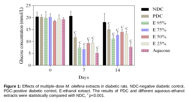
Effects of multiple-dose M. oleifera extracts in diabetic rats
The ethanol (95, 75, 50, and 25 % [v/v]) and aqueous extracts, metformin, significantly reduced the BG levels on days 7 and 14 after overnight fasting (p < 0.001; ). On day 7, the 50 and 25 % (v/v) extracts reduced the BG concentration by 55.35 and 54.57 %, whereas the reduction afforded by the 95 % (v/v) extract was 64.07 % compared with the control. Likewise, the aqueous extract afforded a 60.06 % reduction in BG level compared with the initial value (19.28 ± 0.36 mmol/L).
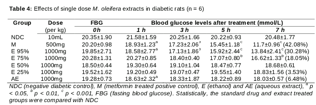
Effects of single-dose M. oleifera extract in diabetic rats
Metformin afforded 42.87 % reduction in hyperglycaemia, compared with the NC, 7 h after administration (). The 95 and 75 % (v/v) ethanol extracts lowered the BG level by 30.28 and 18.05 %, respectively. The 50 and 25 % (v/v) ethanol extracts did not markedly reduce the BG level, whereas the aqueous extract lowered the BG level significantly to only 1 h after administration (p < 0.05).
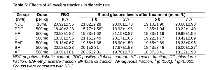
Effects of M. oleifera fractions in diabetic rats
Diabetic rats that received the butanol fraction developed significant (p < 0.01) acute anti-hyperglycaemic activity. No other fraction was effective in this regard ().
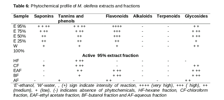
Phytochemical profile
The active butanol fraction and the 95 % (v/v) ethanol extract contained abundant flavonoids, glycosides, tannins, and phenols (). The aqueous extract lacked alkaloids and terpenoids, whereas hexane fraction contained only tannins and phenols.
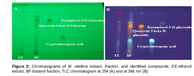
Thin layer chromatogram
shows three prominent spots in the 95 % (v/v) ethanol extract and the butanol fraction. These were identified as quercetin 3-β-D-glucoside, kaempferol 3-O-glucoside, and cryptochlorogenic acid (), with Rf values of 0.658, 0.711, and 0.316, respectively.
Identified compounds and validation results
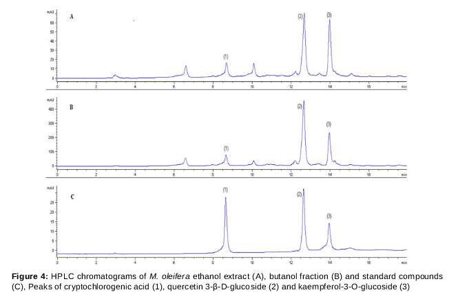
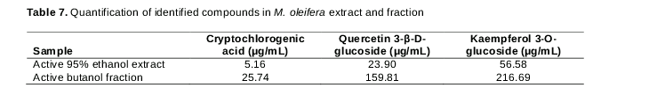
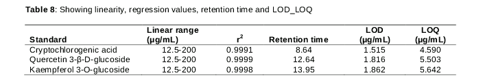
HPLC chromatograms of the active 95 % (v/v) ethanol extract, the butanol fraction, and the standard mixture are shown in . Compound identification was based on the absorption spectra, retention times, and peak heights. Kaempferol-3-O-glucoside was present at high levels in the active 95 % (v/v) ethanol extract and the butanol fraction (56.58 and 216.69 µg/mL, respectively). The potential anti-diabetic cryptochlorogenic acid content of the butanol fraction was 25.74 µg/mL (). The limits of detection for cryptochlorogenic acid, quercetin 3-β-D-glucoside, and kaempferol-3-O-glucoside were 1.515, 1.816 and 1.862 µg/mL, respectively. The limits of quantification were 4.590, 5.503, and 5.642 µg/mL, respectively ().
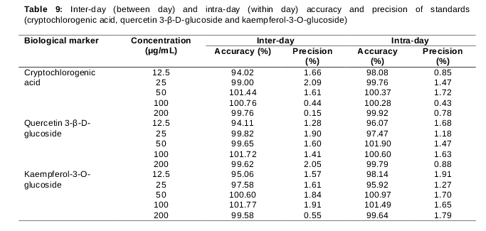
Intra-day accuracy (repeatability) values were from 98.08 - 100.37 % for cryptochlorogenic acid, 96.07 - 101.90 % for quercetin 3-β-D-glucoside, and 95.92 - 101.49 % for Kaempferol-3-O-glucoside. Intra-day precision values were 0.43 - 1.72 % for cryptochlorogenic acid, 0.88 - 1.68 % for quercetin 3-β-D-glucoside, and 1.27 - 1.91 % for kaempferol-3-O-glucoside. Inter-day accuracy values were 94.02 - 101.44 % for cryptochlorogenic acid, 94.11 - 101.72 % for quercetin 3-β-D-glucoside, and 95.06 - 101.77 % for kaempferol-3-O-glucoside. Similarly, the inter-day precision values were 0.44 - 2.09 % for cryptochlorogenic acid, 1.28 - 2.05 % for quercetin 3-β-D-glucoside, and 0.55 - 1.91 % for kaempferol-3-O-glucoside ().
Discussion
Anti-diabetic herbal therapies are very important in the field of ethno-pharmacology. Currently, such therapies are urgently needed to replace oral hypoglycaemic drugs, which are increasingly unable to control all pathological symptoms of diabetes, and are often unavailable in remote areas [14]. We evaluated aqueous ethanol extracts of M. oleifera in normal and streptozotocin-induced diabetic rats to identify possibly relevant compounds.
The rises in BG levels of normal rats after administration of M. oleifera extracts may be attributed to the plant material’s content of a high proportion of carbohydrates. Previous studies showed that the 90 % (v/v) ethanol extract contained 45.43 % carbohydrates [15].
The aqueous-ethanol extracts (95, 75, 50, 25 % [v/v] ethanol and 100 % water) did not exhibit a hypoglycemic effect or affect intraperitoneal GT in normal rats. This lack of effect may be because homeostasis was intact; plant extracts did not interfere with the normal regulation of carbohydrate metabolism [16]. An earlier study [17] also showed no hypoglycaemic effect in normal Wister rats. Data from earlier studies differ somewhat from ours in terms of indications of a hypoglycaemic effect in normal rats; this discrepancy may be attributable to the use of different rat strains, climatic variation in M. oleifera species, and/or the doses given.
We found that the 95 % (v/v) ethanol extract and the butanol fraction thereof were the most active extract and fraction, respectively, exerting very significant acute anti-hyperglycaemic activities in diabetic rats. These effects may be attributable to the expression of glucose transporter-4 (GLUT-4) in the cell membrane, which promotes glucose uptake by skeletal muscle cells and inhibition of glucose synthesis in hepatic cells [18]; skeletal muscle cells constitute 70 % of the total body mass [19]. The results of multiple dosing were in agreement with those of a previous study [20] of the anti-diabetic potential of M. oleifera aqueous ethanol extracts (p < 0.001). The anti-hyperglycaemic effect was greater on day 7 than on day 14, possibly attributable to a reduction in appetite and induction of other anti-diabetic mechanisms.
Initially, we tested various isocratic and gradient systems in terms of optimum separation of compounds using a mobile phase (0.1 % [v/v] formic acid and acetonitrile). The isocratic method did not adequately separate phenolic compounds; only the gradient method produced well-resolved sharp peaks that absorbed at 344 nm.
Linearity was evident over the range of 12.5 - 200 µg/mL for cryptochlorogenic acid, quercetin 3-β-D-glucoside, and kaempferol-3-O-glucoside [r2 = 0.9991 (y = 3.1452x + 3.0369), r2 = 0.9999 (y = 3.1485x - 1.8511), and r2 = 0.9998 (y = 1.1045x + 0.0278) respectively; time vs. peak height in mAU]. The data shown in confirm the validity of the HPLC method in terms of intra-day and inter-day precision, accuracy, and repeatability. TLC also identified isoquercetin, astragalin, and crypto-chlorogenic acid in the 70 % [v/v] ethanol extract of M. oleifera leaves [21].
Conclusion
The anti-hyperglycaemic effect of the M. oleifera 95 % (v/v) ethanol extract and the butanol fraction thereof in diabetic rats may be attributable to cryptochlorogenic acid, quercetin 3-β-D-glucoside, and kaempferol-3-O-glucoside. This extract, and the compounds mentioned, may be alternative treatments for diabetes, and may prompt further drug discovery.
Declarations
Acknowledgement
References
Archives


News Updates